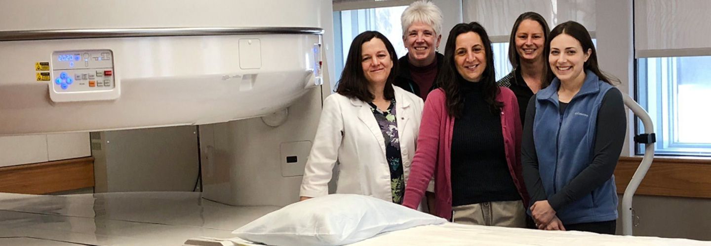
MRI does not use X-rays to create images. Instead, it combines magnetic fields with radiowaves and uses specially designed computers to produce detailed images of internal body structures. While X-rays may be best for showing bones, physicians use MRI to examine "soft" tissue such as muscle, nerves, cartilage, ligaments, tendons, vertebral discs, and various internal organs.
Specialized MRIs
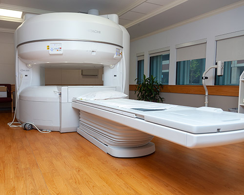
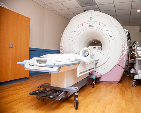
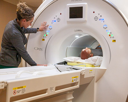
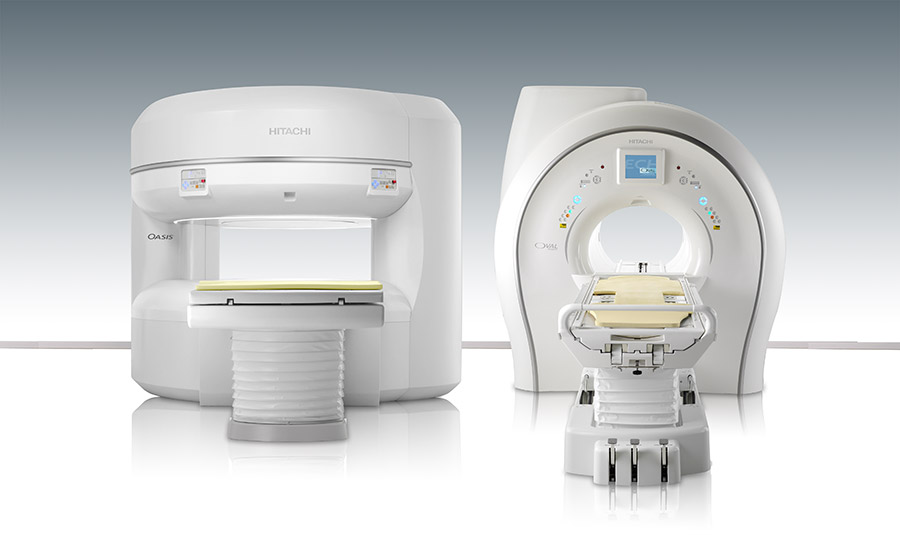
Both CIC locations offer specialized MRI machines to allow more room for patients that need it. The Open MRI unit at CIC Pillsbury features a wide-open design with plenty of space on either side of the patient. The Echelon Oval MRI at Horseshoe Pond features an oval shaped entry that allows much more room above and to the side of patients. Both of these specialized machines are designed to accommodate patients with athletic or large body types, and to alleviate anxious or claustrophobic patients. For more information on how to decide wich MRI will best suite your comfort and needs, please give us a call and we would be happy to help you!
What to Expect
The technologist will escort you to the MRI suite where you will be asked a series of screening questions prior to the MRI exam. Although MRI is an advanced imaging technique, the exam itself is relatively easy and comfortable. You will be asked to lie on a cushioned tab
le. A device called an imaging coil will be placed over or under you. When you are comfortably positioned, the table will move into the open magnet.
From the control area, the technologist will stay in constant contact with you, both visually and through an intercom. You will be asked to remain still while images are obtained. As the exam begins you will hear a variety of muffled thumping or clicking sounds. These sounds are normal and should not be cause for concern. Other than the muted sounds, MRI usually produces no bodily sensations.
Depending on your study, the technologist or nurse may place an intravenous (IV) line through which a MRI contrast agent will be given.
Most MRI exams last between 30 to 60 minutes.
MRI of the head

